Home » Without Label » Bones In Leg Diagram : Tibia Definition Anatomy Facts Britannica : Inside of arm muscle and bone 12 photos of the inside of arm muscle and bone , bone
Bones In Leg Diagram : Tibia Definition Anatomy Facts Britannica : Inside of arm muscle and bone 12 photos of the inside of arm muscle and bone , bone
Bones In Leg Diagram : Tibia Definition Anatomy Facts Britannica : Inside of arm muscle and bone 12 photos of the inside of arm muscle and bone , bone. Related posts of bones leg diagram picture bones and muscles anatomy. Health diagram bone skeleton leg knee science anchor chart human human body. He leg's main function in the human is for locomotion and support of the rest of the body. The smaller lateral bone of the lower leg. The tibia and the fibula, at the top of the ankle joint.
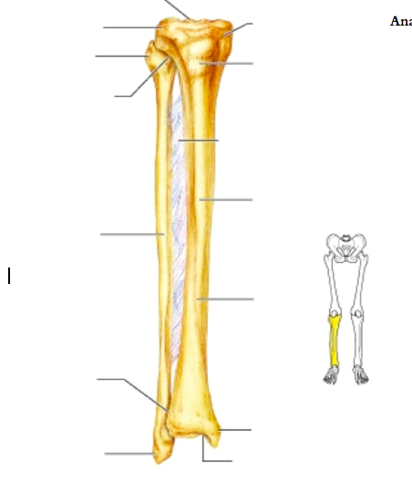
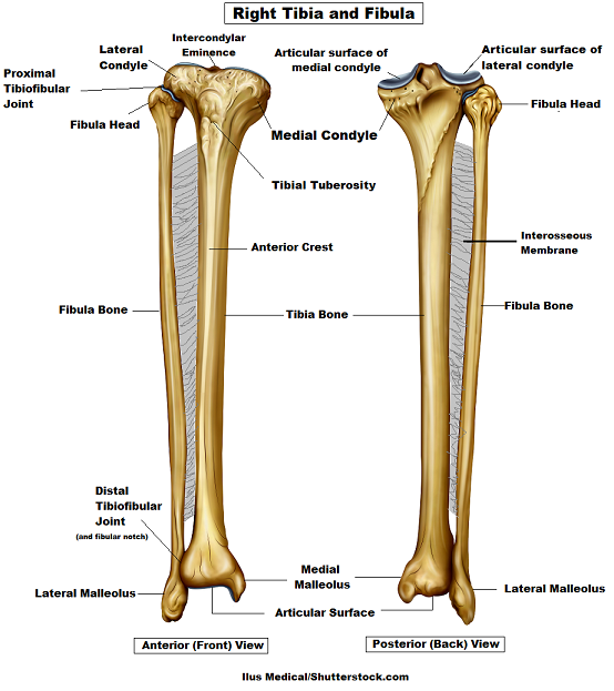
The ligament joining the two bones of the lower leg (tibia and fibula), called the syndesmotic ligament, is injured. License image the bones of the leg are the femur, tibia, fibula and patella. The tibia, commonly known as the 'shin bone', is the largest and most medial of the two.you can palpate its anterior border when you run your finger down the anterior aspect of your leg. The foot bones shown in this diagram are the talus, navicular, cuneiform, cuboid, metatarsals and calcaneus. Posted on april 18, 2019april 18, 2019.
Bones Of The Leg Part 1 Diagram Quizlet from o.quizlet.com Also called the shin bone, the tibia is the longer of the two bones in the. Human leg bone diagram : The diagram of bones in the ankle and foot is given below: Image result for leg bones diagram human leg bone structure your leg bones are the longest and strongest bones in your body. The lower leg extends from the knee to the ankle. The femur, or thighbone, is the longest and largest bone in the human body. The ligament joining the two bones of the lower leg (tibia and fibula), called the syndesmotic ligament, is injured. Its lower end helps create the knee joint.
The tibia, commonly known as the 'shin bone', is the largest and most medial of the two.you can palpate its anterior border when you run your finger down the anterior aspect of your leg.
Master leg and knee anatomy using our topic page. These muscles work together to produce movements such as standing, walking, running, and jumping. These landmarks are the anterior superior iliac spine. This diagram depicts diagram leg bones anatomy.human anatomy diagrams show internal organs, cells, systems, conditions, symptoms and sickness information and/or tips for healthy living. The tibia and the fibula, at the top of the ankle joint. Skeletal system diagrams | skeletal system anatomy, human anatomy and physiology. The femur, or thighbone, is the longest and largest bone within the human physique. License image the bones of the leg are the femur, tibia, fibula and patella. Related posts of bones leg diagram picture. Its decrease finish helps create the knee joint. He leg's main function in the human is for locomotion and support of the rest of the body. The major bones of the leg are the femur (thigh bone), tibia (shin bone), and adjacent fibula, and these are all long bones.the patella (kneecap) is the sesamoid bone in front of the knee.most of the leg skeleton has bony prominences and margins that can be palpated and some serve as anatomical landmarks that define the extent of the leg. The bones of the leg are the femur, tibia, fibula and patella.
The foot bones shown in this diagram are the talus, navicular, cuneiform, cuboid, metatarsals and calcaneus. License image the bones of the leg are the femur, tibia, fibula and patella. The tarsal bones in the foot are located amongst tibia, metatarsal bones, and fibula. Pin on medical websites we like. With different grades of sprains depending on severity.
Lower Leg Bones Diagram Quizlet from o.quizlet.com Master leg and knee anatomy using our topic page. With different grades of sprains depending on severity. The foot bones shown in this diagram are the talus, navicular, cuneiform, cuboid, metatarsals and calcaneus. The diagram of bones in the ankle and foot is given below: The foot bones shown in this diagram are the talus, navicular, cuneiform, cuboid, metatarsals and calcaneus. At the same time, the bones and joints of the leg and foot must be strong enough to support the body's weight while remaining. Pin on medical websites we like. The biceps is attached to the arm bones by.
The proximal portion of the tibia is tibial plateau which acts as a cusp for the knee, the distal portion tapers into the medial malleoli and the concave surface which articulates with the talus at the ankle joint.
Start studying leg bone labeling. The tibia, commonly known as the 'shin bone', is the largest and most medial of the two.you can palpate its anterior border when you run your finger down the anterior aspect of your leg. The biceps is attached to the arm bones by. The tibia and the fibula, at the top of the ankle joint. These muscles work together to produce movements such as standing, walking, running, and jumping. The lower limb contains 30 bones. File human arm bones diagram svg wikipedia. He leg's main function in the human is for locomotion and support of the rest of the body. The human leg consists of 8 bones, 4 per leg. The femur is the single bone of the thigh. These bones are arranged into two major divisions: Ankle & lower leg anatomy. The bones of the leg and foot form part of the appendicular skeleton that supports the many muscles of the lower limbs.
The foot bones shown in this diagram are the talus, navicular, cuneiform, cuboid, metatarsals and calcaneus. Bones in leg diagram color the leg on the left side. The tarsal bones in the foot are located amongst tibia, metatarsal bones, and fibula. The knee joint is the largest joint in the body and is primarily a hinge joint although some sliding and rotation occur. The lower limb contains 30 bones.
Tibia And Fibula Bone Anatomy from www.registerednursern.com The lower limb contains 30 bones. The human leg consists of 8 bones, 4 per leg. Ankle & lower leg anatomy. Master leg and knee anatomy using our topic page. The bones of the leg are the femur, tibia, fibula and patella.the foot bones shown in this diagram are the talus, navicular, cuneiform, cuboid, metatarsals and calcaneus. The leg is specifically the region between the knee joint and the ankle joint. The foot bones shown in this diagram are the talus, navicular, cuneiform, cuboid, metatarsals and calcaneus. The smaller lateral bone of the lower leg.
The biceps is attached to the arm bones by.
Leg bones diagram diagram schematic ideas lower leg muscle diagram blank sketch coloring page antique 1890s medical anatomy diagram leg bones skeleton posted on april 18, 2019april 18, 2019. The bones of the leg and foot form part of the appendicular skeleton that supports the many muscles of the lower limbs. To explain the term in layman's language, it is the heel bone in the skeletal system. This short post will try to cover the dog leg anatomy in detail with labeled diagrams. He leg's main function in the human is for locomotion and support of the rest of the body. The foot bones shown in this diagram are the talus, navicular, cuneiform, cuboid, metatarsals and calcaneus. The diagram of bones in the ankle and foot is given below: The pubis, ischium, and ilium together constitute the pelvis while the thigh bone is the femur. Bone diagram forehead (frontal bone) nose bones (nasals) cheek bone (zygoma) upper jaw (maxilla) lower jaw (mandible) breast bone (sternum) upper arm bone (humerus) lower arm bone (ulna) thigh bone (femur) collar bone (clavicle) toe bones (phalanges) ankle bones (tarsals) kneecap (patella) shin bone These muscles work together to produce movements such as standing, walking, running, and jumping. Pin on medical websites we like. 12 photos of the bones leg diagram picture. The foot bones shown in this diagram are the talus, navicular, cuneiform, cuboid, metatarsals and calcaneus.